Improved image tools for describing diseased and healthy blood vessels

Description (background and methods)
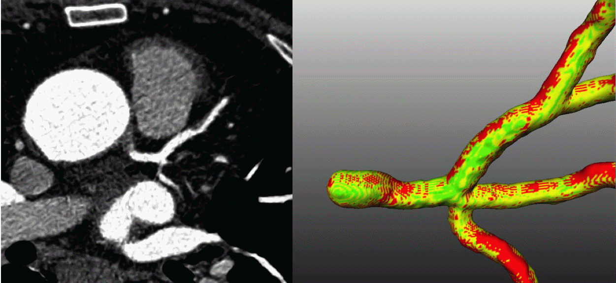
The disastrous effects of atherosclerosis stem from the impaired blood flow through stenoses (constrictions) in the arteries. An increasingly popular way to evaluate the hemodynamic importance of stenoses is to use Computed Tomography Angiography (CTA) combined with segmentation of the arterial lumen and Computational fluid dynamics (CFD) simulations of the blood flow. This project will attempt to answer the following questions:
- How sensitive are computed flow estimates to minor errors in segmentation?
- How large errors are introduced by simplifications in the model?
- Can deep learning improve stenosis quantification or blood flow predictions?
- Can morphometric analysis of the vascular tree identify new risk factors?
In this project, we apply segmentation techniques developed by our group (level sets with coherent propagation) as well as several available alternatives. The resulting lumen geometry is then used for CFD, taking the complex geometry and different rheological models into account.
(Preliminary) results and publications
Andersson MJ, Jägervall K, Eriksson J, Persson A, Granerus G, Wang C, Smedby Ö. How to measure renal artery stenosis - a retrospective comparison of morphological measurement approaches in relation to hemodynamic significance. BMC Medical Imaging 2015;15:42. dx.doi.org/10.1186/s12880-015-0086-8
Medrano-Gràcia P, Ormiston J, Webster M, Beier S, Young A, Ellis C, Wang C, Smedby Ö, Cowan B. A computational atlas of normal coronary artery anatomy. EuroIntervention. 2016;12(7):845-54. dx.doi.org/10.4244/eijv12i7a139
Medrano-Gràcia P, Ormiston J, Webster M, Beier S, Ellis C, Wang C, Smedby Ö, Young A, Cowan B. A Study of Coronary Bifurcation Shape in a Normal Population. Journal of Cardiovascular Translational Research 2017;10(1):82-90. dx.doi.org/10.1007/s12265-016-9720-2
Lidayová K, Frimmel H, Bengtsson E, Smedby Ö. Improved Centerline Tree Detection of Diseased Peripheral Arteries with a Cascading Algorithm for Vascular Segmentation. Journal of Medical Imaging 2017 Apr;4(2):024004. dx.doi.org/10.1117/1.JMI.4.2.024004
Collaborations
- Dept. of Mechanics, KTH
- Dept. of Radiology, Linköping University Hospital
