
Evaluation of speckle tracking based measurements of velocity and deformation
The goal of this study is to evaluate how accurate the speckle tracking algorithms in clinical ultrasound scanner measure tissue velocity and deformation.
Methods
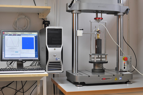
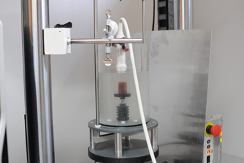
Today, the clinical ultrasound imaging systems are not only used for image rendering but also as a measuring instrument. In the field of cardiology it could be measurements of deformation parameters of the myocardium. These measurements give additional information, but in order to be of clinical value they must have certain accuracy and precision. It is a follow-up study of the earlier published study ”Evaluation of tissue Doppler-based velocity and deformation imaging: a phantom study of ultrasound systems”, where significant differences in measurements between different ultrasound systems were found. An in-house dynamic tissue Doppler phantom was constructed for the previous study. The phantom was based on a commercially available machine for fatigue testing of materials. It was used both to move and compress a tissue mimicking material. The phantom design enabled complex motion and registration of the truly performed motion with high temporal resolution. The same phantom concept will be used in this study. However, another tissue mimicking material with an anatomically-wise more realistic design will be used.
Collaborators
The study is performed in collaboration with the Department of Clinical physiology, Karolinska University Hospital Huddinge.
Contact person
Search words
Ultrasound, speckle tracking, Achilles tendon, evaluation