
Hard x-ray bio imaging
Our research
X-ray imaging technologies are essential for life science, from basic research to clinical practice. Novel modalities such as, e.g., phase-contrast imaging [1], shows promise for higher spatial resolution and improved contrast while still operating with an acceptable dose. Many imaging methods are source limited.
We invented the liquid-metal-jet-anode hard x-ray source [2]. This compact electron-impact microfocus source potentially allows for more than 100´ higher brightness than any competing laboratory x-ray tube. We exploit the unique high spatial coherence of the source for very-high-resolution propagation-based phase-contrast imaging at low dose [3]. Past projects include, e.g., microangiography in mouse, demonstrating detection of sub-10 mm diam vessels. The method was also extended to imaging of intact tumor microvasculature (cf. Fig. 1) [4] as well as to whole-body mouse CT. Current projects are:
Propagation-based phase-contrast imaging
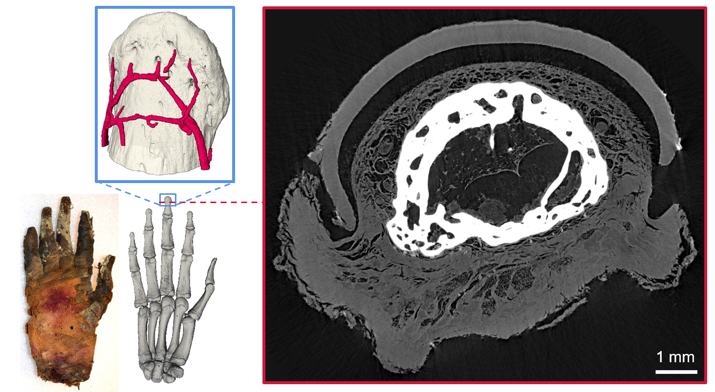
Present work focuses on high-resolution soft-tissue imaging [5,6] and phase retrieval [7]. A recent study concerned imaging of ancient tissue in the form of a 2500 years old mummy hand (see Fig. 2) [8]. Simulations of realistic imaging for medical applications are also developed [9].
X-ray fluorescence tomography
X-ray fluorescence (XRF) is used for 3D molecular imaging. We have developted a laboratory system for high-resolution imaging of small animals [10].
Prospective students
We have master thesis projects available in both phase-contrast imaging and XRF. For more information, please contact Hans Hertz at hans.hertz@biox.kth.se.
References
- See, e.g., F. Pfeiffer et. al, Natute Phys 2, 258 (2006).
- O. Hemberg et al, Appl. Phys. Lett. 83, 1483 (2003).
- T. Tiuhimaa et al, Appl. Phys. Lett. 91, 074104 (2007).
- U. Lundstrom et al, Phys Med Biol. 59, 2801-11 (2014).
- W. Vågberg et al, Sci Rep. 5, 16625 (2015).
- W. Vågberg et al, Sci Rep. 8, 11014 (2018) .
- I. Häggmark et al, Opt. Express 25, 33543 (2017) .
- J. Romell et al, Radiology 289, 670-676 (2018) .
- I. Häggmark et al, IEEE TMI 40, 539-548 (2021) .
- K. Shaker et al, IEEE TMI 39, 3910-3919 (2020) .